Say what? Autofluorescence? You’re telling me parts of my body just light up on their own, like some green cat that’s been bred in a Japanese lab?
Yep, no fooling. Granted, you usually have to be in a dark room and shine a particular wavelength at the tissue in question. And even then, the level of reflection really depends on the tissue. Sometimes it’s only perceptible under a microscope. But, yes, parts of your body light up like that Japanese cat who’s had green fluorescent protein spliced into its genome. Skin and hair tend to both have a low-lying level of autofluorescence. Other tissues like collagen and also many cancerous and precancerous tissues light up under certain wavelengths. In the case of cancers, this can even be used diagnostically [1].
But why am I talking about autofluorescence, except that, like the perpetual children that we are, we love things that are bright and shiny? This is why:
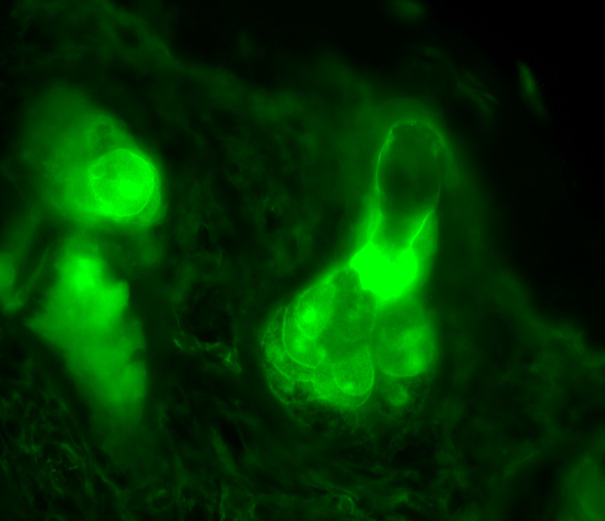
Even though my readers may be most familiar with my genetic research, I also spend some time in the wetlab playing with mice. (I don’t recommend the pass time to any animal lovers such as myself; it’s not pleasant other than some of the husbandry aspects.) Currently, I have a mutant mouse line whose hair follicles are affected by said mutation– an unexpected finding, mind you, but one I’ve elected to study nonetheless. They subsequently lose all their fur, occasionally regrow a little of it back in a form I call the “lion’s mane”, but for the most part are relatively bald. However, when you look at their hair follicles under the epifluorescent scope at 40x mag, this is what you see. BRIGHT GREEN. No stains, no fluorescent tags, no nothing. Nevertheless, a bright green hair follicle emerges into view, carting along some rather hypertrophic-looking sebaceous (oil) glands, which are the well-defined nucleated cells hanging off the follicle in center stage. (This latter characteristic appears to be a secondary result of the mutation.)
Beautiful, no?
But what makes tissues like these fluoresce? Well, a number of molecules, called “fluorophores”, emit light when excited by photons. For instance, coenzymes like flavins and NADH/NADPH fluoresce within the blue/green/yellow range of the visible color spectrum and tend to be good indicators of metabolic activity. These coenzymes only fluoresce in their oxidized forms, meaning that they’re short a proton and have an extra electron with a negative charge on them. A common approach to eliminating autofluorescence is to apply a reducing agent to your preparations which will donate the hydrogen atom and stabilize the molecule. The green fluorescence in the image above is most likely due to one or more of these coenzymes, particularly flavins which glow most strongly in the green range.
Other biomolecules such as porphyrins fluoresce in other parts of the color spectrum, notably within the yellow/red range (600-700nm). In some of my work, I’ve noted red fluorescence of some of the keratin shafts and I’m wondering (though still presently uncertain) whether this isn’t due to a coupling of porphyrins with melanin (pigment) within the hair shaft [2]. I should also mention that melanin fluoresces as well, though in a lower spectral range than porphyrins (360-560nm).
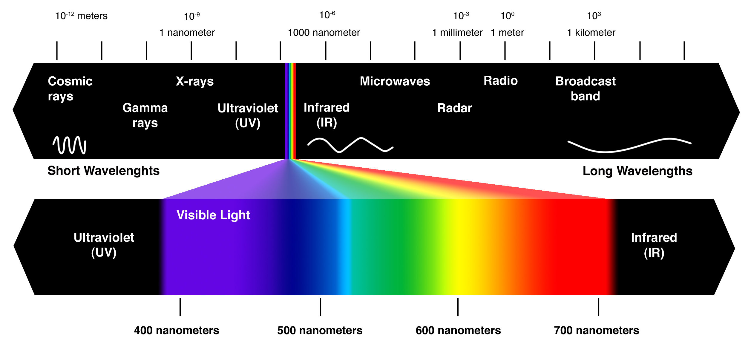
The visible color spectrum, ranging from violet (400nm) to red (700nm). Image borrow from here.
Now I’m no physicist, but I’m going to attempt a simple explanation as to why certain biomolecules fluoresce, so here goes. As I mentioned, most of these fluorophores tend to have a negative charge, which is why using reducing agents that donate a hydrogen atom (i.e., a proton) tend to quash fluorescence. Most molecules, those which do not autofluoresce, do not contain enough internal energy to remain in any vibrational state (a type of electron state) other than its lowest level, S0. Except fluorophores. These little guys are capable of remaining in S1 or even S2 states. When a photon of the appropriate wavelength hits the fluorphore in just the right way, that energy is absorbed, which causes the atom to relax to its lowest level energy state, and a photon is subsequently re-emitted. This is fluorescence [3]. (And for any physicists out there, please feel free to correct or clarify what I’ve written since this is certainly not anywhere near my area of expertise!) For those interested, there are a number of fluorophores in the human body and for a more extensive list please see Wiki’s coverage on the topic with associated references [4].
As I mentioned in the case of cancer, autofluorescence can be used diagnostically. Some researchers have also used this to identify sites and types of infection, since certain bacteria autofluoresce more than others [5]. And in the case of these porphyrin-fluorescent bacteria, treatment with red light can also be used as an effective antibiotic [6].
In the case of my own work, autofluorescence is one of the best ways to study tissues in their most unadulterated postmortem forms– or even premortem forms. In fact, if I wanted to I could take a mouse, give it some anaesthesia to knock it out for awhile, shave and Nair some of its hair off, and put it under a scope to see the hair follicles. All without ever having to euthanize the poor creature. Talk about “in vivo,” eh? The problem with fixing, embedding, cutting, washing, and staining tissues is that, the more you do to it, the more they’re altered from their native forms. Which can ultimately end up affecting results, possibly even falsifying them.
Another important thing for fluorescent studies is knowing WHICH tissues do actually autofluoresce. Look back at that first picture I posted of the green fluorescent hair follicle. Just imagine what I as a scientist would think had I taken that section of skin, stained it with an antibody in order to fluorescently-tag a protein of interest, and then looked under the scope at my results. What I would originally think of as “signal”, indicating the location of whatever protein I’m interested in, would only be the native fluorescence of the tissue itself. What if I tried to draw conclusions from these results? They’d probably be utterly wrong. Especially since my mutant mouse clearly has a very different autofluorescent pattern than the controls.
And being new to hair studies, that’s exactly what happened to me until a kindly senior scientist informed me about what I was actually seeing. All my previous stains now need to be redone on the red color channel since the follicles don’t seem to fluoresce in that color range, except portions of the hair shaft which are easily distinguishable.
It’s a good lesson for caution. It’s also a good lesson in seeking help and advice from those who’ve been around the block more times than you. And it’s an excellent lesson in how friggin’ awesome bright shiny objects are and that the child in us never grows too old for sparkly things.
