Proposing that prenatal ultrasound is a potential teratogen sounds like pseudoscience to most people, I realize this. Hell, when my partner came to me with the idea even I thought he’d gone round the bend. But he really wanted us to review the literature and see if this crazy notion could have any foundation at all. Prenatal ultrasound is after all a growing clinical epidemic and the Rakic team out of Yale had published a paper back in 2006 that found changes in the brains of prenatally exposed mice, so why not? [1]
With my reservations in hand, I started scanning the literature. At first all the early safety studies from the ’70s and ’80s seemed to say the same thing: “No deleterious changes following exposure.” Again and again I came across the same types of studies, many epidemiological. After awhile and in frustration, I set the searches aside because the literature at first glance really seemed to suggest that ultrasound was relatively safe, and the occasions in which differences did arise, such as low birth weight, they eventually righted themselves. So not much to go on.
After a nice hiatus investigating other things, I ran across an article that showed that when osteoblasts, fibroblasts, and monocytes were subjected to ultrasound in vitro (in a petri dish), exposure stimulated the production of interleukin-8, basic fibroblast growth factor (bFGF), and vascular endothelial growth factor (VEGF), each of which acts as growth factors on these and nearby cells [2]. That same group later found that osteoblasts under similar levels of exposure release nitric oxide and prostaglandin E2, both of which are known to have growth-inducing effects on bone [3].
Even though we’re talking bone and not my primary focus, brain, these results truly intrigued me. Unfortunately, one can’t be certain from such an experiment that the same would be found in vivo (in life) as opposed to a petri dish and whether such effects occur across cell types. But the fact that the effect was real ignited my curiosity and I was primed to continue my searches into the biophysical mechanisms of ultrasound. It hasn’t been easy because the research is spread across numerous professional disciplines, but here’s a basic rundown of my literary travels:
Ultrasound is exactly that, “ultra” and “sound”. It’s an auditory waveform that occurs above the upper limit for human hearing. Like all sounds– and as is implied by the term “waveform”– this flow of sound pressure occurs in waves, called compression and expansion half-cycles. All that simply means is that the materials that are hit by this sound undergo alternations of a compression of their materials followed by an expansion, kind of like an accordion. This is one of the things that makes ultrasound potentially dangerous, dependent on the level of intensity of the soundwave and the duration of exposure.
The act of bubble or cavity formation within a liquid upon ultrasound exposure is called cavitation. The term cavitation is particularly used to refer to the implosion of a gaseous bubble under pressure within a liquid, although this is more specifically known as transient cavitation. Stable cavitation is the formation of bubbles which remain relatively stable in the liquid medium. In pure water, bubbles form at more than 1,000 atmospheres of pressure, which is to say that if you were to take pure water and subject it to even the highest intensities of diagnostic or prenatal ultrasound, bubbles would not form because the intensity is far too low. But not so for biological tissues. As occurs with almost every solid within a liquid (e.g., cells), small gaseous pockets hide within crevices. The pre-existence of such bubbles essentially lowers the threshold for cavitation within biologic tissue compared to pure water because gaseous cavities, though extremely small, are already present. When subjected to the force of ultrasound, these bubbles undergo expansion with the expansion half cycles and compression with the compression half cycles. Just picture taking a balloon, blowing it up, then sucking in a little of the air and seeing it contract a little bit, and repeat this procedure over and over again. In this analogy you are the ultrasound wave, the force driving expansion and contraction. The main problem is that with each subsequent cycle, the bubble compresses less and less, so over time the size of it grows until at which point, much like when that balloon is filled till it pops, the surface area of the bubble can no longer withstand the pressure. In the case of a bubble in liquid, it implodes and the surrounding water rushes in, meets with the gases once trapped in the bubble, triggering a violent chemical reaction which produces an extraordinary local rise in temperature– a temperature close to that of the surface of the sun [4]. Thankfully, the rapid cooling rates of the surrounding medium (on the order of 109 ⁰C·s-1) virtually assure that for a single occurrence the temperature of surrounding tissues rises insignificantly. The problem arises when transient cavitation occurs more frequently in a local tissue such that the rapidity of cooling is not as efficient which can cause thermally-induced tissue damage.
These little bubbles can also cause other problems aside from temperature increase. As you might imagine, when a bubble implodes it can create a considerable force of pressure in the surrounding medium such that nearby cells may be hit with high pressure water jets. This can also occur, though in a gentler fashion, by stable cavitation in which the bubble doesn’t implode but merely oscillates next to or within a cell, creating a variety of forces and pressures against the outer membrane and within the cell itself. In a worst case scenario, the jets of water may fatally damage the membrane and internal structures of cells, leading to cell death. In a less deadly scenario, the water jet may poke transient holes through the outer membrane of the cell which allows into the cell many communicating molecules such as sodium, calcium, and various proteins. This inward rush of calcium, for instance, can activate many downstream pathways involved in cell repair, cell growth, and even alter cell-to-cell communication, such as in the case of neurons. Did you know in fact that ultrasound is used transcranially (across the skull) to elicit activity of target neurons? [5]
So what harm can a little calcium do? Well, if it doesn’t kill off the cell through calcium cytotoxicity, the fact that many of these extracellular molecules are normally used in a controlled way for cell-to-cell communication essentially feeds cells the wrong messages and could feasibly alter the development of cells permanently, especially concerning stem cells, progenitors, or immature cells which may pass down these alterations through further generations.
Previous in vitro studies have also found that ultrasound is capable of promoting chromosomal breakages in DNA, although there isn’t currently evidence to suggest the same severity is found in vivo within diagnostic and prenatal intensity ranges so this may be an effect particular to cell cultures [6]. But it does beg the question what ultrasound exposure may be doing to the cellular contents within exposed cells. Poking transient holes through membranes, jostling the cell’s internal contents, disturbing its general physiology, and if exposure occurs for a long enough duration, the greater the likelihood of cavitation, deadly jets of water, and thermally-induced damage.
Think of it: You subject cells to increased water pressure, and if the duration is long enough the threshold for transient cavitation may be reached. The potential for ultrasound’s teratogenicity is considerable and, in this scientist’s opinion, research has not been thorough enough to rule out possible dangers. Consider the ultrasound parties which are becoming all the rage or the keepsake ultrasounds parents are having done to “start the family photo album early”,– or what about the doppler ultrasound fetal heart rate monitors that parents can use unsupervised in their own homes on a daily basis? And that’s not even addressing the issues of poor technician education or the staggeringly high rates of malfunctioning ultrasound equipment used in everyday clinical practice [7, 8]. Ultrasound regulations are dismal in this country and may unknowingly be turning a useful tool into a teratogen.
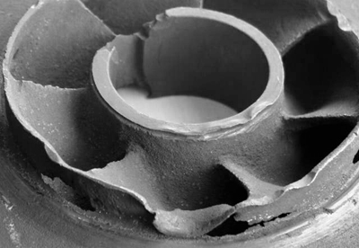
I want you to think about this: The image below is a pump which has been degraded by transient cavitation stimulated by ultrasonic waves. If you have enough force, this is what occurs to metal. Just think what could be happening to a developing fetus. Admittedly, this image is meant to scare you. And while the intensity levels and duration of exposure which a fetus endures are by no means comparable to what this water pump has gone through, it’s the same basic physics based upon cavitational and noncavitational effects.
We truly need to temper our enthusiasm for ultrasound photos of our babies and reassess whether this tool is as safe as we think it is. As a clinical tool it is extraordinarily useful, and not unlike X-rays, it has its purposes. But it isn’t just a photograph.
As a parting gift, below is a list of some of the many uses of ultrasound in medicine, manufacturing, and research. It’s been borrowed from one of our in-press publications (Williams & Casanova, 2013). I hope it might start the cogwheels turning.
- Diagnostic sonography providing structural imaging, including prenatal ultrasound;
- The ablation of target tissue, i.e., during neurosurgery or tumor removal, and the breakdown of calculi i.e. kidney stones or gallstones;
- Transcranial ultrasonic stimulation, similar to transcranial magnetic stimulation (TMS);
- Vasodilation, providing better visualization of the vasculature during cardiovascular procedures;
- Targeted drug delivery, utilizing focused ultrasound to stimulate greater tissue more permeability, e.g., the blood-brain barrier, skin, etc.;
- Wound healing;, e.g., bone fractures and ulcers;
- Bactericidal properties when synergized with antibiotics;
- Elastography, in which ultrasound is used to determine the elasticity of a given organ which can help discern the overall health of that organ;
- Transmembrane delivery of products into target cells, e.g., nonviral genes or nutrients;
- Acoustophoresis: the use of ultrasound on an ionic medium to create an electric charge;
- The purification of agricultural products;
- Heat transfer in liquids for production of substances such as ethanol;
- The purification of metals;
- Manipulation and characterization of particles in the bio- and physical sciences;
- The testing of metals, plastics, aerospace composites, wood, concrete, cement, etc. in manufacturing in order to measure thickness and locate flaws within the material.

I was hoping to see you post something on this topic ever since I noticed a mention of the potential dangers of ultra-sound in a twitter comment on the right-hand side of the page.
Good for you. I have to read the above fully and carefully and that must wait until I have more time.
Meanwhile, I hope many thousands of people shall read your views on this topic and circulate them to their friends and family.
I hope this does circulate so that awareness raises on this topic. It’s in dire need of reinvestigation.
I am really pleased you wrote this and are calling for reinvestigation. My son jumped a mile when they turned on the ultrasound at my 20 week scan and then proceeded to hide. He refused to budge and they couldn’t get all the measurements! As sensible little foetus I think!
This was16 years ago and at the time women were having far fewer scans than they are now, and it was supposed to be safe. I wasn’t so sure after my son’s reaction, so declined a further scan to check for measurements that they didn’t get the first time. As far as I was concerned they saw that everything was basically fine, I didn’t need to know his leg measurements and what sex he was.
Now though it means that I will always wonder if that single scan was enough to make his Ehlers Danlos type 3 and his ASD worse than that of either of his parents.
I recall an article that discussed the level of movement of the fetus responding to ultrasound exposure (i.e., increased). I’ll have to see if I can dig that up somewhere… it’s been awhile…
Would you (or, may I, with your help and advice,) link or re-post this in another science forum, citing your (and Dr. Casanova’s ?) text/ideas/thoughts in whole or in part? When you have a chance, I’d like to know what you’d think about that.
Thank you for another excellent treatment of an important topic.
Feel free to link or re-post, proximity. 🙂
how do you feel about the new ultrasound technique of ablation of fat? as a sort of refined liposuction without the need for surgery….
That’s hard to say. Without knowing too much about the technique, I would assume that a high intensity is used in the procedure. Truthfully, using a tool which hasn’t been thoroughly studied is concerning in any of its uses on biologic tissues. I would be curious as to the width of the sound beam and how much tissue, beyond that of adipose, is being exposed.
Pingback: The Need for Further Studies on the Safety of Prenatal Ultrasound | Cortical Chauvinism·
Pingback: La necesidad de estudios adicionales sobre la seguridad de la ultrasonografia o ecografía prenatal | Cortical Chauvinism·
a contributor whose user-name is “Mud”, registered and signed in to contribute a very thoughtful and well-written comment in support of your thesis here–at that site where I’d re-posted the text and, which site I won’t bother to mention again here. Suffice it to say that the supporting post was as welcome as it was insightful and when I read it, I thought, I wish I’d said, written, those same things as well.
To “Mud”, (should he or she chance to see this) : thank you for that effort. No other commenter has shown enough insight to do as much there.
What kind of pump is this? Knowing what kind of pump this is and what is it used for would help with the trying to show “similarities”. Also the power behind the U/S’s they have in the medical field and what the power behind this “pump” would be also helpful. Along with the time durations.
Hi, Corrin. Sorry for the delay in reply. I can’t honestly say I know the precise intensity, frequency, and duration on the industrial pump. It’s one of a number of images available showing what long-term exposure to US can do on equipment. However, you have as much information at your disposal as I do (Google), if you’re interested in comparing. I would recommend just looking up intensity and frequency of US in industrial pumps. (Sorry for not readily providing you the info, but work is horrifically busy at the moment.) As I said in the blog post though, the duration of exposure alone makes the severity of what has occurred to this pump poorly comparable to the exposure a fetus receives. However, the severity of exposure wasn’t really the point but to stress that the same mechanisms which are creating an ultrasound image or audio are the ones which, over long periods of exposure, eventually erode metals like in the pump image above. Even though the effects on the industrial pump are much more severe, these effects overall lie on a continuum. The big question is whether we have fully and satisfactorily addressed safety on the prenatal US end. And the point of the blog wasn’t to say that ultrasounds as they’re currently used are definitely unsafe. The point was to say that we don’t know for certain they are, especially given how poorly regulated their use. And that’s something we need to find out and, if necessary, place tighter regulations upon.
Thanks so much for the post. I always avoided ultrasounds like fire and I never really had an explanation except for the concern of what these “sound waves” are actually doing to my fetus. I’m happy to read a more scientific point of view.
My only problem reading this article is a technical one – it’s really hard reading white on black. It’s not very eye friendly. It would be great if you could change the color scheme.
Thanks again!
Hi, Alina. Just as a quick note, since I can totally understand the sensory issue regarding the website having sensory issues myself, I’ve linked to the website, Readability, in the lefthand sidebar which automatically simplifies websites to a plainer text formatting. It was very recently suggested to me and so I’m trying to let readers know about it for exactly the reasons you state. I would change the design except that 1) I have sensitivity towards light bright screens and find the white on black easier to handle, and 2) have run across more people with a similar sensitivity vs the preference for black on white. So I hope that helps somewhat.
Hi, everyone. Sorry for the delay in replies; I’ve been battling a bad case of sinusitis and am now back at work but having to catch up on some experiments. I want to reply thoughtfully so I will do so hopefully over the weekend.
I had a high risk pregnancy in my mid-forties, carrying twins. Frequent ultrasounds were done – to establish pregnancy, to check for growth, to rule out birth defects, etc. A level two ultrasound and 3d imagery were included in this package. The end result? I have boy/girl twins with autism. It could have been genetic – my children were donor embryos. It could have been me – more toxins in an older body, being released as I depleted old stores of nutrients, muscle and body fat towards the end of pregnancy when I could not eat enough to keep from losing weight. Could it have been the fillings? Or the in vitro processes and cryopreservation techniques? No one can say. While correlation does not prove causation, it is interesting to see that the incidence of autism and the frequency of prenatal ultrasound usage follow a similar trend.
Yes, it definitely raises questions about safety. Regarding autism there may be several factors affecting its development and need not be any single one. Thanks for your story and your neutral skepticism.
I’ve had 4 children & 5 ultrasounds and with each one, I’ve been more reluctant, more concerned about the effects. I hate that I did them at all, especially for my second son. The ultrasound went on for so long and I just wanted to tell them to stop, but didn’t have the courage to stand up for myself, or him. He’s 8 now and still has speech issues, as well as ADHD tendencies and anger management problems. I can’t help wondering how much that ultrasound is to blame.
Yes, it seems to be very hard for many people to protest against doctors’ instructions. On the one hand, they do know more about medicine, anatomy, and physiology usually than the patient. On the other hand, current medicine isn’t perfect either and mistakes are made. I hope that ultrasound isn’t one of them, but we still need to do more research to determine whether that’s the case or not. My partner has some preliminary work with collaborators on an animal model which looks promising. If the remainder of the data follows the same trend, then it should be a very informative publication, which I’ll certainly be covering in future here on SoaC. So please stay tuned!
This is a great article which raises some interesting and valid points! I’d really like to be able to share it, but it contains an ableist term: “crazy”. If you wouldn’t mind altering your post to use a different word, I’d love to pass this along! Thanks.
Thanks for the compliments, erthemama. 🙂
As to your request, truthfully I feel conflicted. I genuinely understand the avoidance of certain terms because they may paint a group of people in an unpleasant, unfair, or inaccurate light. For instance, I try my best to avoid using terms of “pathology” when I can– especially things like “disease” or “disorder”. So I truly get where you’re coming from.
On the other hand, sometimes I feel people might place so much importance on words that we forget they’re just words. Words can truly hurt, there’s no doubt about that. But they’re also an imperfect inaccurate medium with which to convey thought, and a label will almost always be inaccurate– perhaps even insulting– in some circumstance or another. They will never be perfect. And in this day and age of political correctness, it’s probably better to strive for a golden medium than to sway to either extreme. Just my opinion though, take it as such.
It’s really hard not to want to change other people. I know, we spend a lifetime trying to do it. However, I did write the word “crazy” and while it may not have been the best choice of words, it was what appealed to me at the time.
So to be fair, I don’t think I’m going to change the word. However, I do promise you that I will make a point to consider its use much more carefully in future, especially when I may be putting people out if I do use it. I’ll think much more carefully about, not just what it means to me, but all the things it may mean to other people. And I’ll ask myself, “Is this what I want to convey?”
Now, is that a better win for you, the fact you are inspiring me to change my behavior, rather than to simply have me change a single word and not give much further thought to it?
I’ll be interested to hear what you think. Take care.
and now,
“Stethoscope set to be supplanted by new technology
Heart experts predict pocket-sized ultrasound machines will improve diagnostic accuracy and reduce complications”
The Guardian (London) reports: http://www.theguardian.com/society/2014/jan/24/stethoscope-supplanted-new-technology-ultrasound#start-of-comments
Patients, buy your own stethoscope now–while you still can find one and bring it with you to your appointed visits with your GP or specialist because science is letting us down again over the matter of patient-safety versus doctors’ technological conveniences.
Hey, proximity. Welcome back. 🙂
On the one hand, ultrasounds used to detect heart abnormalities could definitely be life saving. They would certainly offer a much more refined means of detection. On the other hand, the fact that complications from ultrasound have been so poorly studied really leaves one anxious and wondering what new problems could arise from this kind of use. My reaction to this is a combination “Brilliant!” and “Oh shit…” Thanks for sharing!
After reading this article I am so glad my midwife did not trust ultrasound use, on the unborn. Even back then I had second thoughts about it, even without her input.
There are some conditions that can be diagnosed and treated in the third trimester, especially closer to delivery, that can improve mother/infant safety. So I would ‘t necessarily throw the metaphorical baby out with the bathwater. A single short ultrasound in later pregnancy can be wise. But ultrasound in earlier trimesters seems unnecessary and risky.