Connective tissue is a fibrous cell-sparse network that helps to connect, support, bind, and separate neighboring tissues from one another. It exists in and around every organ of the body. Probably the most recognizable forms of connective tissue are bones (calcified), tendons, ligaments, cartilage, and fats. One major component of connective tissue is the extracellular matrix (ECM), which is composed of various molecules (e.g., proteins) that give structural and communicative support to nearby cells.
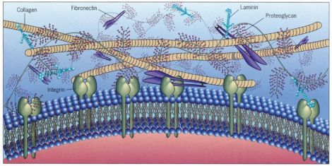
Common examples of the ECM include elastins and collagens, the latter the most abundant protein in the human body and which is highly implicated in different forms of Ehlers-Danlos syndrome (EDS) [1]. Mutations in the collagen 3 gene, for instance, are associated with the vascular form of EDS, while collagen 5 mutations underlie many forms of classic EDS. Some of our recent work has focused on the potential overlap between EDS and the autism spectrum. While I won’t go into much detail at the moment (stay tuned for upcoming publications from our lab), there is nevertheless growing interest in EDS, its association with neurodevelopmental conditions like autism and ADHD, and how collagen deficits may play a role in those conditions.
Unlike tendons, for instance, the human brain doesn’t house what one would call “connective tissue” in the traditional sense. Whenever one stains for the ECM in a slice of brain tissue, the intensity of that stain is, shall we say, underwhelming compared to other organs like the skin? However, ECM in the brain does nevertheless maintain an extensive network that, like other forms of connective tissue, both supports and separates neighboring tissues from one another.
As an example, the ECM supports the growth and establishment of neuronal axons and dendrites and also helps maintain synaptic connections [2]. Interestingly, the local ECM must be degraded and remodeled to allow for synaptic plasticity– a process that’s dependent on neural activity [3]. Scientists have also found that the ECM forms compartments surrounding neurons that help to limit the diffusion of the excitatory neurotransmitter, glutamate– allowing refinement of those signals between nearby neurons [4]. Without such barriers, glutamate would be allowed to reach more distant neurons, substantially altering patterns of cell communication.
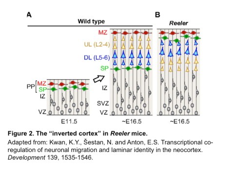
Much earlier in development, during embryogenesis, some types of collagen such as collagen 5 are also expressed in the germinal regions of the brain in areas that are actively producing neurons [5]. While it’s uncertain exactly what collagen 5 may be doing during neurogenesis, it’s known that other forms of collagen (e.g., collagen 4) are expressed in these same regions during early development and seem to be important for neuronal maturation [6]. Other forms of ECM proteins, such as reelin, are vital for proper migration of neurons into the developing cortex. Reeler mice (genetically-modified mice that have mutations in the reelin gene) have what is known as an “inverted cortex,” in which the neurons migrate and form cortical layers in an outside-in fashion, rather than the normal inside-out manner (figure below).
Although we don’t yet know if collagen deficiencies seen in EDS and related hypermobility spectrum disorders (HSD) have measurable effects on early brain development, the links between EDS/HSD and various neurodevelopmental conditions like autism suggest collagen may play a larger role than is currently recognized. We also don’t know if these effects are primarily limited to the early prenatal period, given collagen’s expression within the germinal zone of the developing brain, or if it plays other roles in the mature brain that are currently unrecognized.
Unfortunately, the ECM within the brain has received minimal attention despite hints at its importance. Though there are undoubtedly a number of reasons for this, the fact that connective tissue within the brain is comparatively sparse and must be studied at the molecular level rather than under a microscope has essentially resulted in scientific “out of sight, out of mind.” But as scientists continue to root out their blindspots, and as interest from patient communities changes, I think the roles of ECM in brain development will continue to receive more and more attention.

This link interests me. I have a son with severe autism. Now my daughter is being evaluated for EDS. She has hypermobility.
Just curious: Did your son experience any regression?
Pingback: Connective Tissue & the Brain | EDS and Chronic Pain News & Info·
Yes! I am a school psychologist and I have EDS. I’ve been curious about this. I am finding I am experiencing a lot of symptoms of Autism, but it isn’t making such an impact to a disordered level. I’ve been diagnosed with ADHD and I wonder if I’ve always just been subdiagnostic autistic. I also have known many students with hypermobility and Autism. I think there is likely an EDS link there and imagine that connective tissue problems in the brain result in disruptions in brain processing. Also, I know that when I’m experiencing pain (which is almost all the time these days), I am more easily overstimulated and only want to engage in my special interests that are particularly soothing because it is too taxing to engage in other things. It is all very fascinating.
Hi, Jaymie. You may well be a Broader Autism Phenotype (BAP). I suspect there’s a sizeable minority of the EDS population that are BAP or autistic.